Microscopy Collaboration Highlight

Researchers combined three unique types of microscopy to track how a protein named clathrin triggers cell membrane bending. The research team essentially filmed live cells internalizing their own membrane using fluorescence on a nanoscale.They found that clathrin has an unexpected amount of plasticity when pinching off small portions of the cell membrane. Their work was published in the Jan. 29, 2018, issue of Nature Communications.
What is the impact?
The visualization provides a better understanding of the mechanism that allows cells to internalize beneficial nutrients as well as fight viruses. A greater understanding of how cells internalize material will help biotechnology companies such as SAB Biotherapeutics develop more treatments for influenza and other diseases.
What is the significance of this outcome?
The collaboration was a unique team science effort that brought together three microscopy teams utilizing imaging tools acquired with NSF EPSCoR support to visualize clathrin-mediated cell membrane bending. Teams from South Dakota State University and the South Dakota School of Mines worked with National Institutues of Health scientists Justin Taraska and Kem Sochacki on the project.
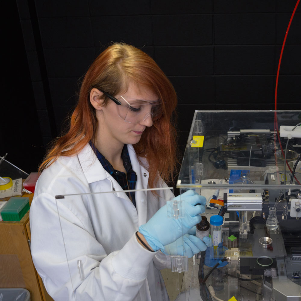
The researchers used a laser beam to differentiate horizontal and vertical orientations and chemically froze the cells at various stages to capture high-resolution images of membrane bending. The research answered a fundamental ‘how does the cell work’ kind of question that has radiating impacts on a multitude of disease processes and important physiological functions in plants, animals and humans. The research collaboration was a true team effort to see live cells internalizing their own membrane.
Read more about this research in the 2018 SD EPSCoR Summer Newsletter.
 National Science Foundation RII Track-1 Project:Expanding Research, Education and Innovation in South Dakota